Introduction: Understanding Cherry Angiomas
Cherry angiomas, also known as senile angiomas or Campbell de Morgan spots, are among the most common benign vascular proliferations encountered in dermatological practice. These lesions are composed of dilated capillaries and postcapillary venules within the papillary dermis, typically presenting as small, bright red papules on the trunk and extremities.
While most cherry angiomas are small, asymptomatic, and cosmetically inconsequential, nodular or pedunculated variants can pose clinical challenges. These larger lesions may bleed spontaneously, increase in size over time, or cause significant cosmetic concern—particularly when located in visible areas such as the scalp.
Clinical Significance
Nodular cherry angiomas represent a deeper and more compact vascular proliferation compared to typical cherry angiomas. Their tendency to bleed with minimal trauma and potential for growth makes them ideal candidates for procedural intervention rather than observation.
Case Presentation
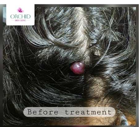
A 35-year-old female patient presented to the outpatient dermatology clinic with a solitary, dome-shaped, bright red nodular lesion on the scalp. The lesion had been present for approximately 8 months with gradual increase in size, causing considerable anxiety due to its vascular appearance and tendency to bleed during hair combing.
Clinical Features
On examination, the following characteristics were noted:
- Location: Scalp region
- Morphology: Well-circumscribed, smooth, erythematous nodular papule
- Size: Approximately 6-7 mm in diameter
- Consistency: Soft and compressible
- Symptomatology: Occasional bleeding with minimal trauma
- Associated features: No pain, ulceration, or surrounding inflammation
- Systemic: No similar lesions elsewhere on the body
The patient reported significant distress related to the lesion's appearance and its tendency to bleed when combing hair, impacting her quality of life and necessitating intervention.
Diagnostic Approach
The diagnosis of nodular cherry angioma was established clinically based on characteristic morphological features and clinical behavior. The diagnostic process involved:
Clinical Assessment
Key diagnostic features supporting the clinical diagnosis included:
- Typical coloration: Bright red appearance characteristic of vascular lesions
- Compressibility: Lesion blanched partially with pressure, confirming vascular nature
- Surface characteristics: Smooth surface without keratosis or pigmentation
- Bleeding tendency: Consistent with friable vascular tissue
Differential Diagnosis
Several conditions were considered and systematically excluded:
- Pyogenic granuloma: Excluded due to absence of rapid growth and marked friability
- Angiokeratoma: Ruled out by lack of keratotic surface changes
- Amelanotic melanoma: Not consistent with irregular borders or atypical features
- Hemangioma: Clinical course and morphology more consistent with cherry angioma
Diagnostic Confidence
Given the classic clinical presentation with typical morphology, color, and behavior, the diagnosis was made with high confidence. Pre-treatment biopsy was deemed unnecessary, allowing for immediate therapeutic intervention.
Understanding the Pathophysiology
Cherry angiomas arise from benign proliferation of dilated capillaries within the superficial dermis. While the exact etiology remains incompletely understood, several factors are believed to contribute to their development:
Proposed Mechanisms
- Age-related changes: Increasing prevalence with advancing age, present in over 75% of individuals by age 75
- Hormonal influences: Potential role of hormonal factors, particularly during pregnancy and hormonal transitions
- Genetic predisposition: Familial clustering suggests hereditary component
- Angiogenic dysregulation: Altered expression of vascular endothelial growth factor (VEGF) and other angiogenic mediators
- Environmental factors: Possible association with chemical exposure, though evidence remains limited
Nodular Variants
Nodular cherry angiomas represent a subset characterized by:
- Deeper dermal extension with more compact vascular proliferation
- Larger vessel caliber and increased vascularity
- Greater propensity for bleeding and trauma-related complications
- Negligible potential for spontaneous regression
Treatment Approach: Radiofrequency Electrocautery
After discussing treatment options and obtaining informed consent, radiofrequency (RF) electrocautery was selected as the optimal approach for this nodular cherry angioma. This technique offers several advantages for scalp lesions, particularly precision, excellent hemostasis, and superior cosmetic outcomes.
Surgical Technique
Step-by-Step Procedural Approach:
- Preparation: Strict aseptic precautions with cleaning of the operative field using antiseptic solution
- Anesthesia: Local infiltration with 2% lignocaine administered around the lesion base
- Lesion engagement: Gentle grasping of the lesion with surgical forceps
- RF application: Application of radiofrequency current in cut-coagulation mode at the lesion base
- Complete removal: Systematic coagulation ensuring complete lesion removal
- Hemostasis: Immediate and complete hemostasis achieved through RF coagulation
- Wound inspection: Verification of clean coagulated base with no residual lesion
Advantages of RF Electrocautery
Radiofrequency electrocautery offers distinct benefits for vascular lesions:
- Precision: Targeted tissue destruction with minimal collateral damage
- Superior hemostasis: Simultaneous cutting and coagulation
- Speed: Rapid procedure completion (typically under 10 minutes)
- Cosmetic outcomes: Minimal scarring when performed correctly
- Office-based: No need for operating room or general anesthesia
- Cost-effective: Lower cost compared to laser therapies
Post-operative Care
A comprehensive post-operative protocol was instituted to optimize healing and prevent complications:
- Topical antibiotic: Fusidic acid cream applied twice daily for 5 days to prevent secondary bacterial infection
- Systemic anti-inflammatory: Tablet Enzoflam for 2-3 days for pain and inflammation control
- Wound care instructions: Gentle scalp hygiene, avoiding aggressive manipulation of the treatment site
- Activity modification: Avoidance of hair combing over the treated area for 48-72 hours
- Follow-up schedule: Planned review at 1 week and 4 weeks post-procedure
Treatment Outcome
The procedure was well-tolerated with negligible intraoperative bleeding. Immediate post-procedure examination revealed a clean, coagulated base with excellent cosmetic appearance. The patient reported high satisfaction with the outcome, with complete resolution of bleeding episodes and cosmetic concerns.
Excellent Results
Early follow-up demonstrated successful lesion clearance with no evidence of recurrence or complications. The treatment site showed minimal scarring, validating the choice of RF electrocautery for this scalp lesion.
First Visit
Well-circumscribed, erythematous nodular cherry angioma measuring 6-7mm on scalp with tendency to bleed with minimal trauma.
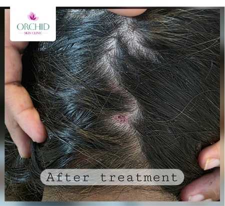
Four Weeks Post-Treatment
Complete lesion clearance with excellent cosmetic outcome and minimal scarring following radiofrequency electrocautery.
Treatment with RF electrocautery provided complete resolution with no recurrence at extended follow-up.
Alternative Treatment Modalities
While RF electrocautery was selected for this case, several other effective treatment options exist for cherry angiomas. The choice depends on lesion characteristics, location, number, and patient preferences:
Surgical Options
- Standard electrocautery: Using needle or loop electrodes for lesion destruction
- Surgical excision: Complete removal with primary closure (reserved for large or atypical lesions)
- Shave excision: Tangential removal at skin level with base cauterization
Non-surgical Approaches
- Cryotherapy: Liquid nitrogen application (effective for smaller, superficial lesions)
- Pulsed dye laser (PDL): Selective photothermolysis of vascular structures
- Nd:YAG laser: Deeper penetration for larger vascular lesions
- Intense pulsed light (IPL): Broad-spectrum light targeting hemoglobin
Selection Rationale
RF electrocautery was specifically chosen for this case due to:
- Scalp location requiring excellent hemostasis
- Nodular morphology benefiting from depth control
- Cost-effectiveness and immediate availability
- Superior outcomes reported for pedunculated/nodular variants
- Single-session treatment capability
Clinical Implications and Key Learning Points
This case highlights several important clinical principles in the management of nodular cherry angiomas:
Diagnostic Considerations
- Clinical diagnosis suffices: Typical cherry angiomas can be diagnosed confidently based on morphology alone
- Know when to biopsy: Atypical features warrant histopathological confirmation
- Differential diagnosis matters: Always consider amelanotic melanoma in rapidly growing vascular lesions
Treatment Principles
- Patient-centered approach: Consider psychological impact and quality of life
- Technique selection: Choose modality based on lesion characteristics and available resources
- Optimize hemostasis: Critical for scalp lesions due to rich vascularity
- Prevent recurrence: Ensure complete base coagulation to minimize recurrence risk
Prognostic Factors
- Low recurrence: Adequate coagulation typically results in permanent clearance
- New lesion development: Patients may develop additional cherry angiomas over time
- Scarring potential: Minimal with proper technique and post-operative care
Clinical Pearl
For scalp cherry angiomas, radiofrequency electrocautery offers the optimal balance of efficacy, safety, and cosmetic outcome. The technique's inherent hemostatic properties are particularly valuable in this highly vascular location.
Conclusion and Future Directions
This case demonstrates the safety and efficacy of radiofrequency electrocautery for nodular cherry angiomas, particularly in challenging locations such as the scalp. The technique provided excellent lesion clearance with superior cosmetic results and high patient satisfaction.
Key takeaway messages include:
- Cherry angiomas, though benign, merit treatment when symptomatic or cosmetically concerning
- Radiofrequency electrocautery represents an excellent office-based treatment option
- Proper technique and post-operative care are essential for optimal outcomes
- Patient counseling about the benign nature reduces anxiety
- Early intervention prevents bleeding episodes and patient distress
As our understanding of angiogenic mechanisms continues to evolve, future research may reveal targeted preventive strategies. However, current treatment modalities, particularly RF electrocautery, provide reliable and effective management for symptomatic lesions.